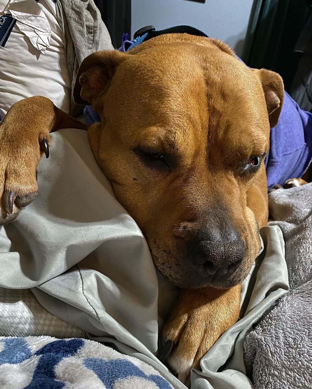
Have you noticed something different about your dog’s eyelid, eye, or the whole side of its face? You have had your dog for a couple of years and everything has been alright… You go on walkies, play catch, and say what a good boy he is… But!
Something started to change in and around your dog’s eye lately, making you wonder if your doggo had an injury or developed an allergy.
There is something different with the upper eyelid… even the pupils seem strange. So, what could it be?
Well, if you take your dog to a vet, she or he can examine your dog and make a diagnosis: Horner’s syndrome in dogs. We say ‘in dogs’ because Horner’s syndrome can affect people as well.
People often get scared and think of the worst when they hear the word “syndrome”. But, don’t start panicking, and take a deep breath…
It is not a life-threatening health condition. Alright, now that you know your dog cannot die from Horner’s syndrome, let’s see what can be done to help the dog get rid of it, and what actually happens.
What Is Horner’s Syndrome In Dogs?

Let us start with the definition of a syndrome: it is a group of symptoms and other medical signs that happen at the same time, and are connected to each other, implying there is a disease or a health condition present in the body.
So, Horner’s syndrome can sound bad because people often connect it with more serious syndromes (e.g., Swimmer puppy syndrome) that can actually change the life of the affected dog.
However, Horner’s syndrome in dogs is a condition that does not change the life of a dog, it is not life-threatening, and in most cases, it resolves on its own without any medical help.
Horner’s syndrome affects a dog’s face muscles (facial muscles) and eyes, including eye muscles and pupils. The change happens because of the abnormality in the innervation of the mentioned body parts and tissues.
The Innervation
Just like humans, dogs have an autonomic nervous system consisting of two “parts” that control our entire body. Sometimes, we are conscious of the actions of the nervous system, and sometimes, we are not.
These two systems are called:
- Parasympathetic nervous system
- Sympathetic nervous system
Both systems are made of nerves that go throughout the body, reaching the organs and tissues from head to toe. The biggest difference is the fact that the parasympathetic nervous system relaxes us after a stressful experience.
It is also called automatic because it controls body functions that we cannot consciously control, for example, digestion, heart rate, and sweating.
The sympathetic nervous system is the opposite — it makes us ready to “fight or flight”. It alarms the entire body, and gets it ready to face danger, whatever that might be. It doesn’t have to be connected with danger alone.
An over-excited dog that wags its tail too much and too fast (and possibly develops happy tail syndrome) also has a sympathetic nervous system, which is dominant at that moment. Once the excitement goes away, the parasympathetic system takes control and calms the dog.
Which System Is Affected In Horner’s Syndrome?
Horner’s syndrome in dogs (and in humans) is a result of nerve damage, injury, or abnormal innervation due to the chemical imbalance of the sympathetic nervous system.
There are six cranial nerves that innervate the eye and its surrounding tissues. They are under control by both the sympathetic and the parasympathetic nervous systems, but only the nerve branches under sympathetic control are affected.
This means that the pupil stays constricted because the parasympathetic nervous system does that to the pupil, but it cannot dilate. The sympathetic nervous system dilates the pupils, but in this case, it cannot because there is sympathetic nerve dysfunction.
All of this tells us that the clinical signs of Horner’s syndrome in dogs are a result of damage, lesion, or some other kind of nerve dysfunction in the sympathetic autonomic nervous system.
What Are The Symptoms Of Horner’s Syndrome In Dogs?

So, we understand that the problem occurs because of the problems with the innervation coming from the sympathetic nervous system. What exactly are those symptoms?
These are the things you can notice on your dog’s face:
- Constricted pupils (miosis)
- The affected eye looks sunken (enophthalmos)
- The upper eyelid is droopy (ptosis)
- A red and enlarged third eyelid on the affected side (conjunctival hyperemia, or eyelid prolapse)
Basically, what you will see if your dog has Horner’s syndrome is that the affected eye looks as if it has a sunken eyeball, its third eyelid looks red and larger than a normal eyelid, and the pupil looks like a small dot — as if it is a bright sunny day.
In most cases, Horner syndrome in dogs is unilateral, which means it only affects one side of the face. However, in some cases of Horner syndrome, it can be bilateral — affecting both sides.
You can test your dog’s pupil dilation by shining a flashlight into your dog’s eye. Start from the corner of the eye and pass over the eye with the light. A normal pupil should react to the light and shrink. When the light passes, it should dilate.
With Horner’s syndrome in dogs, this does not happen. The pupil of the affected eye stays the same no matter if the light changes.
If your dog has three of four clinical symptoms upon a DVM examination, the diagnosis is Horner’s syndrome. Isolated clinical signs without the other three or two do not mean that a dog has this health condition.
It could be something completely different that has nothing to do with the neurologic issue. For example, your dog can have dry eyes, allergies, or some other eye problem.
Diagnostic Methods
First of all, it is the pet owner who notices the first signs that point to Horner syndrome in dogs. Naturally, to understand what is happening or to find an underlying cause, we have to take the dog to the veterinarian, a local vet clinic, or a hospital.
Your veterinarian can then do the first step in making a diagnosis — a physical examination. This means that a vet has to thoroughly examine your dog to see the changes or abnormalities in or on your dog’s body.
They will check the eyes, nose, mouth, teeth, and ears. The vet will pay attention to many things at the same time — the color of the mentioned body part, along with its shape and smell. They’ll also pay attention if there are any foreign bodies or changes in the coat.
Sometimes, a vet can order diagnostic tests to be done, like blood work or an X-ray. This is in case the symptoms are either inconclusive or a vet suspects some other issue happening at the same time. Besides typical X-rays, a vet can also do:
- Advanced imaging (CT or MRI)
- Examination of the ear
- Thoracic radiographs
- Pharmacologic tests
All of these tests are done in order to not only make a diagnosis, but to locate the most affected area. The better we understand a disease or a health issue, the easier and more successful the fight against it will be.
A thorough examination is the key to the correct diagnosis, the most effective treatment, and consequently the easiest and quickest recovery.
However, when it comes to nerve lesions and other innervation problems, the treatment is not that simple. It takes time and patience.
But first, let’s take a look at some of the most common causes of Horner’s syndrome in dogs.
What Can Cause Horner’s Syndrome In Dogs?

In essence, the cause of all the changes that happen in Horner’s syndrome is nerve damage. This damage can be caused by numerous reasons:
- Idiopathic (unknown)
- Trauma
- Infection
- Neoplastic (cancer)
- Iatrogenic
The reason why so many things can damage the sympathetic nerves and cause Horner’s syndrome in dogs lies in the fact that the sympathetic pathway is very long. The longer the nerve is, the more chance it has to be damaged somewhere on its pathway.
To explain it more clearly, we have to divide lesions according to three locations — central, preganglionic, and postganglionic.
Postganglionic
This lesion localization is also called a third-order lesion. As with any other lesion, the cause can be idiopathic, traumatic, neoplastic, or a “simple” ear infection.
The nerve that innervates the eyes also has branches that go into the ear canal. These parts are all connected, and as a result, otitis media or interna (infection of the middle ear or inner ear) can cause lesions of the nerves that innervate the pupil, eyelids, and eye muscles.
Besides ear infections and ear diseases, postganglionic lesions can also happen either without an apparent reason (idiopathic) or as a result of:
- Neuroblastoma
- Carotid body paraganglioma
- Iatrogenic (caused by medical examination or treatment)
Central
This is the segment of the sympathetic pathway that starts at the hypothalamus. This is the place where the cranial nerves exit the brain and continue their way through the brainstem and down the spinal cord.
This lesion localization is called a first-order lesion, and in most cases, it includes brain injuries or neoplastic changes. However, these lesions can happen anywhere in the spinal cord.
The lesions can have the following etiology:
- Idiopathic
- Traumatic
- Infections
- Fibrocartilaginous embolism — cervical
- Neoplastic
Traumatic injuries, neoplasms, embolisms, or infections cause swelling of the tissue (or abnormal growth), which causes physical damage to the nerves, causing total or partial nerve palsy.
Nerve palsy is a lack of function (of a nerve).
Preganglionic
The other name for preganglionic lesions is second-order lesions. The preganglionic segment refers to the pathway that the nerves take as they leave the spinal cord and enter the chest cavity.
The nerves leave the spinal cord at the second thoracic vertebra, and they stretch on both sides of the chest cavity. These nerves innervate the chest, but they also stretch upwards and innervate the neck.
Lesions in this segment can often happen because of trauma from pulling the leash too hard or when we lift the dog by its forelegs. The nerves can stretch too much and suffer a traumatic injury. A traumatic injury can happen during the birthing process or as a result of a bite wound as well.
Besides trauma, these are other reasons for preganglionic lesions:
- Idiopathic
- Iatrogenic (e.g., thoracic surgery)
- Infectious (tick paralysis)
- Neoplastic (lymphomas or the PNST of the vagus nerve)
Other Reasons
Because Horner’s syndrome in dogs is a neurological health issue, the reasons behind the lesions and nerve palsy can be numerous. Besides the mentioned reasons, doctors have found a possible connection between Horner’s syndrome and some other health issues.
For example, there is a higher incidence of Horner’s syndrome in dogs that suffer from hypothyroidism or those suffering from glaucoma.
A connection has also been found (or noticed) between Horner’s syndrome and particular dog breeds. We have to mention that Horner’s syndrome can affect any dog breed, no matter the size, origin, or sex.
However, these dog breeds are more prone to Horner’s syndrome:
- Golden Retrievers
- Collies
- Shetland Sheepdogs
- Labrador Retrievers
- Doberman Pinschers
- Weimaraners
- Cocker Spaniels
This doesn’t mean that every Weimaraner or Doberan Pinscher is going to have Horner’s syndrome, especially not when it comes to traumatic etiology. These dog breeds are simply more prone to idiopathic Horner’s syndrome.
How To Treat Horner’s Syndrome

The treatment of Horner’s syndrome in dogs depends on the etiology. If we know the reason, then we can treat the reason and deal with the syndrome, too.
For example, if the reason behind Horner’s syndrome is a tumor — the treatment focuses on the removal of the tumor whether it is benign or malignant.
If the reason is an infection, then the focus is on the infection. Veterinarians can use eye drops with phenylephrine to subdue the infection and decrease the size of the inflamed third eyelid. But, the eye drops are dealing with symptoms and not the underlying cause — the infection.
So, we have to find out what is causing the infection — a virus or bacteria. After that, we need to know which one and how to treat it.
This is the case with known reasons for Horner’s syndrome. But, what happens when we don’t know the reason, or when we label the syndrome as “idiopathic”?
Unfortunately, there are no specific ways to treat idiopathic Horner’s syndrome. This is because we don’t know what to treat. So, the best we can do is help the dog feel as comfortable as possible.
The Prognosis
Fortunately, in most cases, idiopathic Horner’s syndrome resolves on its own. It seems like a passing nerve dysfunction that goes away after a few months. There is no need for any specific treatment because it is purely a cosmetic issue and not a health issue.
That’s why being patient is the key because signs of Horner’s syndrome can last for more than six months sometimes.
If the reason behind this health condition is an infection or a tumor, or some other pathological cause, the prognosis depends on the severity of the cause as well as the localization of the lesion.
See Next: Why Is My Dog Acting Weird All Of A Sudden?
The Conclusion
Horner’s syndrome in dogs is a health issue that can happen to any dog breed. In most cases, it affects dogs that are older than two years. However, it can happen at any age. Sometimes, a dog starts exhibiting Horner’s syndrome symptoms from birth.
This usually happens because of trauma during the birthing process. So, it can happen from day one to day twenty-one.
Thankfully, this is a condition that often goes by on its own. It does take some time to pass, but it does pass in the end without any health consequences for the dog. This is the case of idiopathic Horner’s syndrome (the one we don’t know the reason behind).
However, because Horner’s syndrome etiology can be quite different, and with pathological roots, it is important to take your dog to the vet as soon as you notice these symptoms: droopy eyelid, constricted pupil, enlarged third eyelid, etc.
Early diagnosis and early localization of the nerve lesion lead to a better prognosis and final outcome.


